


Technical Data
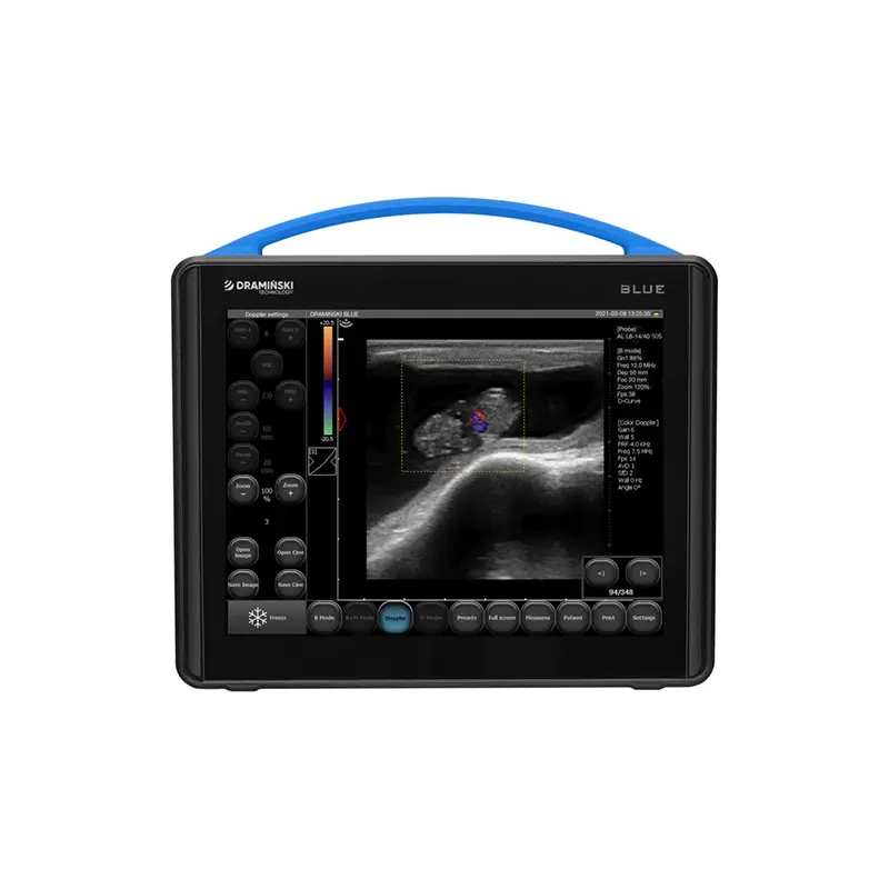
Imaging modes | B Mode B+B Mode 4B Mode M Mode B+M Mode Color Doppler Power Doppler (PDI) Pulse Wave Doppler (PWD) |
Frequency | range: 1-14 MHz (depends on probe) |
Probes | Backfat 2-5 MHz Convex 1-6 MHz Convex 2-5 MHz Convex 2-8 MHz Hockey stick 6-14 MHz Abdominal linear 4-12 MHz Abdominal linear 6-14 MHz Microconvex 4-9 MHz OPU 4-9 MHz Rectal linear 4-9 MHz Rectal convex 2-8 MHz Rectal microconvex (P-probe) 4-9 MHz |
Dynamic Focus | Yes |
Image management | Freeze Zoom 60 – 300% Full screen Invert left-right and up-down Saving images Saving cine loops |
Imaging adjustment | Dynamic range Frame averaging Vi-Probe D-Curve LuciD Compound Imaging (CI) Line density Field of view (FOV) TGC |
Presets | Depend on probe: SDFT, DDFT, Horse SL, Horse foot, Horse back, Horse eye Dog cardio, Dog abdominal, Cat cardio, Cat abdominal FAST |
Postprocessing | Yes (LuciD) |
Measurements | Basic: Distance, Follicle, Area, Ellipse Area, Grid, Stenosis, Volume Obstetric package: aging tables for different species Cardiology package: Heart Rate (HR), LA/Ao, Left Ventricle function (LV), Left Ventricle Volume: V Simpson’s LVAM – LVAP method, V Simpson’s Single plane method, V Bullet method Doppler: Resistance Index (RI), Pulsation Index (PI), Peak Systolic Velocity (PSV), End-Diastolic Velocity (EDV), Average Velocity (AVG) Doppler Point Acceleration Time (AT) Heart Rate (HR) Blood Flow Area % |
Workflow tools | Battery check when OFF Hidden active fields Easy distance measurement Precise measurements with magnifying glass |
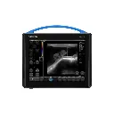
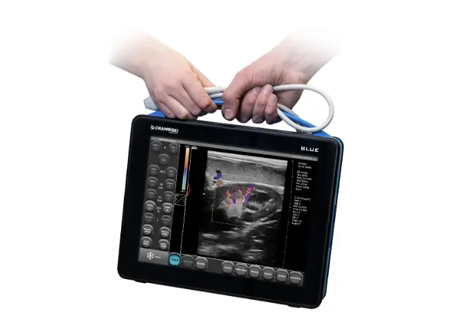
Display | 12″ LCD LED 500 cd/m2 1024 x 768 px |
Control panel | Capacitive touchscreen |
Memory | 30 GB |
Exported file format | PNG, AVI, MP4, MOV, DICOM 3.0 |
Exporting data method | To external flash drive via USB |
Multimedia ports | 1 x USB 3.0, 1 x USB 2.0, 2 x LAN, 1 x HDMI |
WiFi connection to mobile devices | No |
Power supply | Power adapter and built-in battery |
Battery operating time | Up to 2 h 30 mins. |
Battery charging time | Up to 4 h |
Booting time | About 30 s |
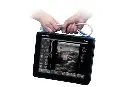
Dimensions | Width 31 cm, Height 28 cm, Depth 6.5 cm |
Weight (with probe and battery) | 4 kg |
Ingress Protection (IP) rating | IP30 |
Accessories
- Wheeled high stand
- Wheeled stand for tendons examination
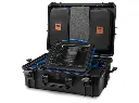
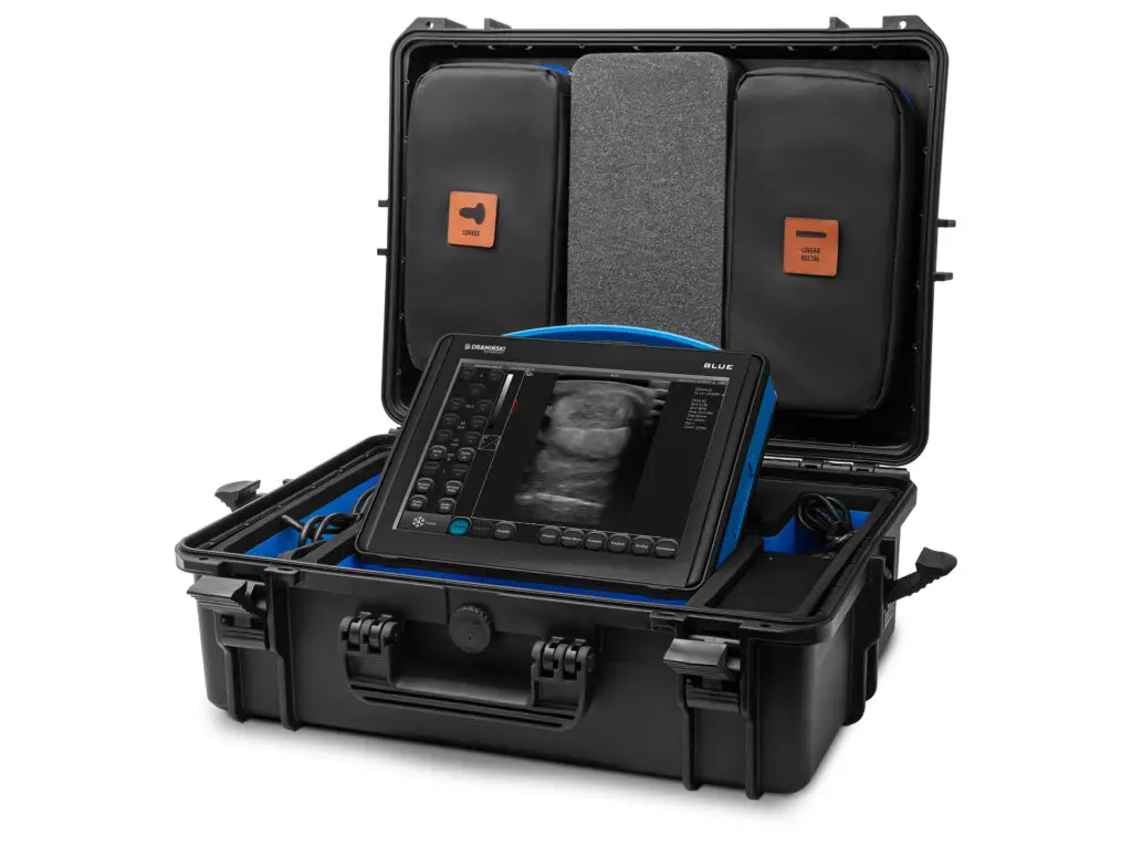
- Transport case with wheels
- OPU
- Extension and T-holder for rectal linear or rectal convex probe
- Shade cover
- Suspenders
- Waist belt
- Standoff
- Wi-Fi Antenna